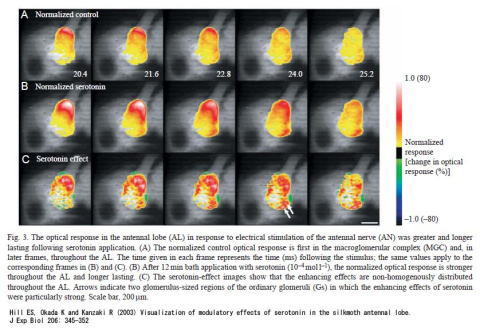
The brain is componsed of a large number of neurons and signal processing occurs in neuronal populations. A method to simultaneously record the activity of many neurons is imaging. One imaging method consists of staining the neural tissue with a dye associating with the cell membranes and being capable of detecting the transmembrane voltage. In this example, the brain was stained with the voltage-sensitive fluorescent dye RH414, whose changes in fluorescence are correlated with the transmembrane voltage. Spatiotemporal activity patterns in the antennal lobe in response to electrical stimulation of the antennal nerve are shown with a time resolution of 0.6ms.
The time series of images demonstrates that serotonin application enhances activation in the antennal lobe. This is physiological evidence that closely matches the fact that serotonin enhances behavioural sensitivity to pheromone.
Functional imaging in the antennal lobe